Back Of Skull And Neck Anatomy / David Weinstock Nkt On Instagram My New Client Was Referred To Me By My Friend The Dentist For Tmjd O Head Muscles Face Muscles Anatomy Human Muscle Anatomy / This vessel also supplies more superficial parts and structures of the head and neck.
Back Of Skull And Neck Anatomy / David Weinstock Nkt On Instagram My New Client Was Referred To Me By My Friend The Dentist For Tmjd O Head Muscles Face Muscles Anatomy Human Muscle Anatomy / This vessel also supplies more superficial parts and structures of the head and neck.. One reason for shooting neck pain at the base of your skull is a pinched nerve in your upper spine. Think of it like a jigsaw puzzle, all the pieces fit in together and are required to get the full picture as to how it works. The cervical spine has 7 stacked bones called vertebrae, labeled c1 through c7. Man, woman head, brain nose, mouth, foot, ear, lips vector illustration. It involves the upper cervical spine, facet joints, muscles, tendons, ligaments, and nerves.
Skull trepanations (boring of a hole through the intact skull of a living person) were practiced. A pinched nerve in your cervical spine is called cervical radiculopathy. Its clavicular head originates from the medial third of the clavicle, while its sternal head arises from the manubrium of sternum . Man, woman head, brain nose, mouth, foot, ear, lips vector illustration. It involves the upper cervical spine, facet joints, muscles, tendons, ligaments, and nerves.
Neck Muscles And Other Soft Tissues from embed.widencdn.net Occipital neuralgia is caused due to irritation or injury to the occipital nerve. While classified as peripheral nerves, the motor cell body resides in the anterior horn of the spinal cord. Bone of back of skull. In other words, there is a muscle on the forehead (frontalis) and one on the back of the. Irritation or injury to any one of these structures can result in pain at the base of the skull. One reason for shooting neck pain at the base of your skull is a pinched nerve in your upper spine. The occipital glands (lymphoglandulæ occipitales), one to three in nu ber, are placed on the back of the head close to the margin of the trapezius and resting on the insertion of the semispinalis capitis.their afferent vessels drain the occipital region of the scalp, while their efferents pass to the superior deep cervical glands. See anatomy of the head and neck stock video clips.
The majority of these nerves control the functions of the upper extremities and allow you to feel your arms, shoulder, and back of your head.
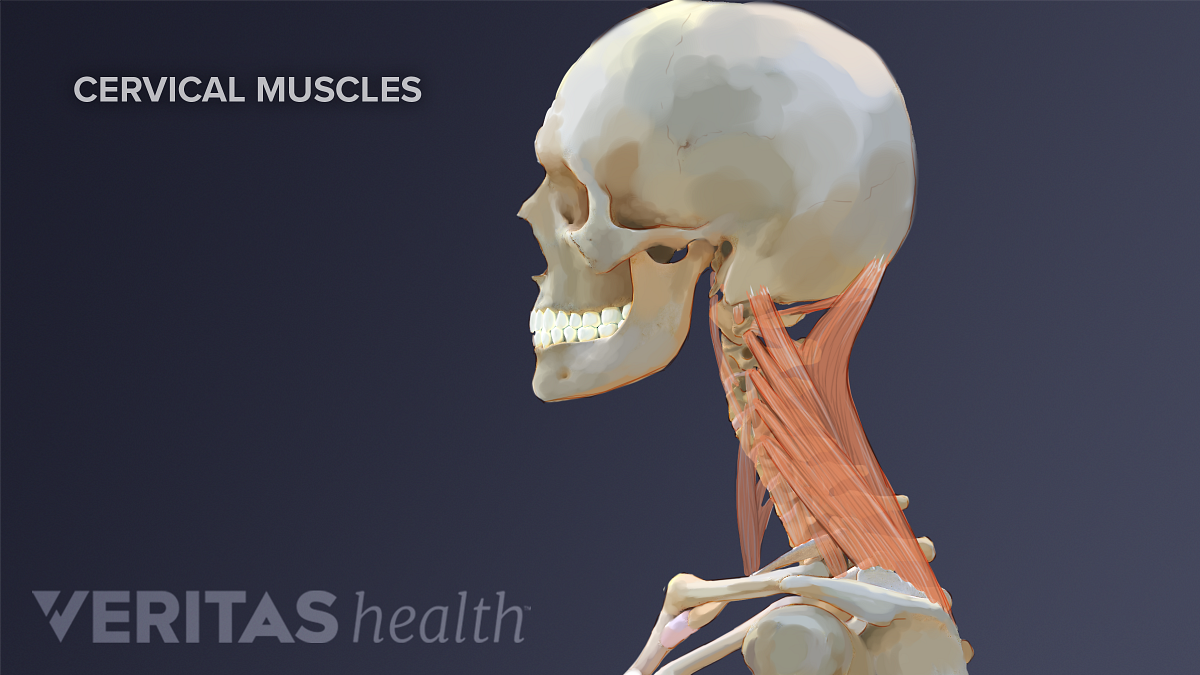
Occipital neuralgia is caused due to irritation or injury to the occipital nerve. The neck begins at the base of the skull and connects to the thoracic spine (the upper back). The neck is connected to the upper back through a series of seven vertebral segments. The cervical spine has 7 stacked bones called vertebrae, labeled c1 through c7. Irritation or injury to any one of these structures can result in pain at the base of the skull. Anatomy the base of the skull is a complex area. Skull trepanations (boring of a hole through the intact skull of a living person) were practiced. Ullrich says that the inflammation in the facet joints in the cervical spine between the shoulders and base of the head causes pain at the back of the head. It consists of two major parts: The head rests on the top part of the vertebral column, with the skull joining at c1 (the first cervical vertebra known as the atlas). The occipital glands (lymphoglandulæ occipitales), one to three in nu ber, are placed on the back of the head close to the margin of the trapezius and resting on the insertion of the semispinalis capitis.their afferent vessels drain the occipital region of the scalp, while their efferents pass to the superior deep cervical glands. Neck anatomy nerves picture there are 8 spinal nerves that originate from the cervical spine. Causes of neck pain and how to manage the pain in basic terms, the neck (cervical spine) joins the shoulders and chest to the head.
These nerves conduct motor and sensory information via efferent and afferent fibers, respectively, to and from the central nervous system. While classified as peripheral nerves, the motor cell body resides in the anterior horn of the spinal cord. The motion of the muscles of the neck are divided into four. Houman body parts flat line icons set. The neck begins at the base of the skull and connects to the thoracic spine (the upper back).
Cervicogenic Headache Causes And Risk Factors from embed.widencdn.net The neck contains seven of. The skull is a strong, bony capsule that rests on the neck and encloses the brain. The nerves of the head and neck include the most vital and important organs of the nervous system — the brain and spinal cord — as well as the organs of the special senses. Bone of back of skull. The neck begins at the base of the skull and connects to the thoracic spine (the upper back). Anatomy of the skull and bones of cranium on medical illustrations. The motion of the muscles of the neck are divided into four. While classified as peripheral nerves, the motor cell body resides in the anterior horn of the spinal cord.
The nerves of the head and neck include the most vital and important organs of the nervous system — the brain and spinal cord — as well as the organs of the special senses.
The skeletal section of the head and neck forms the top part of the axial skeleton and is made up of the skull, hyoid bone, auditory ossicles, and cervical spine. Included in this review are the cervica. The occipital bone is a bone that covers the back of your head; The heads come together and ascend diagonally to insert onto the mastoid process of the temporal bone . Houman body parts flat line icons set. An area called the occiput. The majority of these nerves control the functions of the upper extremities and allow you to feel your arms, shoulder, and back of your head. Causes of neck pain and how to manage the pain in basic terms, the neck (cervical spine) joins the shoulders and chest to the head. The neck begins at the base of the skull and connects to the thoracic spine (the upper back). The neck is connected to the upper back through a series of seven vertebral segments. In addition, in this region we also find the major cranial and spinal nerves that connect the central nervous system to the organs, skin, and muscles of the head and neck. Irritation or injury to any one of these structures can result in pain at the base of the skull. The cervical spine, your neck, is a complex structure making up the first region of the spinal column starting immediately below the skull and ending at the first thoracic vertebra.
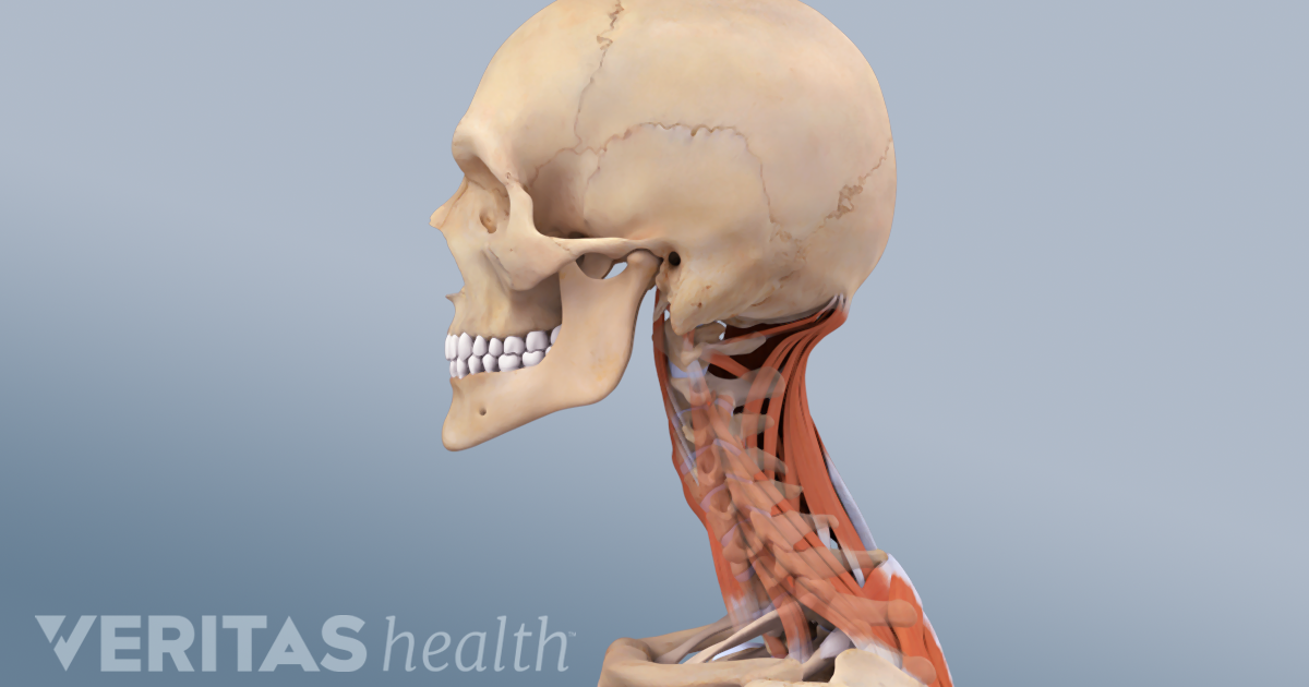
Arthritis sufferers often have headaches in the back of the head and neck. The large, complex muscles of the neck and back move the head, shoulders, and vertebral column. It consists of two major parts: Man, woman head, brain nose, mouth, foot, ear, lips vector illustration. The occipital bone is the only bone in your head that connects with your cervical spine (neck).
Head And Neck Anatomy from fpnotebook.com There are eight pairs of cervical nerves, denoted c1 to c8. The anatomy of the head and neck of the human body, including the bones, muscles, blood vessels, nerves, glands, nose, mouth, and throat. The skeletal section of the head and neck forms the top part of the axial skeleton and is made up of the skull, hyoid bone, auditory ossicles, and cervical spine. The occipital bone is a bone that covers the back of your head; Occipital neuralgia is caused due to irritation or injury to the occipital nerve. Muscle head anatomy vocal organ diagram female neck anatomy neck wireframe head neck human anatomy head artery anatomy face pharynx vector neck degree head anatomy 3d. From the sides and the back of the neck, the splenius capitis inserts onto the head region, and the splenius cervicis extends onto the cervical region. The top of the cervical spine connects to the skull, and the bottom connects to the upper back at about shoulder level.
Each nerve provides sensation to a specific area of the body called a dermatome.
The orbicularis oris is a circular muscle that moves the lips, and the orbicularis oculi is a circular muscle that closes the eye.the occipitofrontalis muscle moves up the scalp and eyebrows.the muscle has a frontal belly and an occipital (near the occipital bone on the posterior part of the skull) belly. Anatomy the base of the skull is a complex area. The neck contains seven of. Ullrich says that the inflammation in the facet joints in the cervical spine between the shoulders and base of the head causes pain at the back of the head. The neck is connected to the upper back through a series of seven vertebral segments. Each nerve provides sensation to a specific area of the body called a dermatome. Causes of neck pain and how to manage the pain in basic terms, the neck (cervical spine) joins the shoulders and chest to the head. The neck is one of the most complex and intricate structures in our body and includes the spinal cord, which sends messages from the brain to the rest of the body. Muscle head anatomy vocal organ diagram female neck anatomy neck wireframe head neck human anatomy head artery anatomy face pharynx vector neck degree head anatomy 3d. The occipital bone surrounds a large opening known as the foramen magnum. From the sides and the back of the neck, the splenius capitis inserts onto the head region, and the splenius cervicis extends onto the cervical region. The anatomy of the head and neck of the human body, including the bones, muscles, blood vessels, nerves, glands, nose, mouth, and throat. The nerves of the head and neck include the most vital and important organs of the nervous system — the brain and spinal cord — as well as the organs of the special senses.


0 Komentar